


Long-term success and survival of post-endodontic restorations without posts
The authors of this paper report a lack of clinical trials on direct/indirect restorations placed on endodontically treated teeth where a post has not been used. They report a high rate of catastrophic failures in teeth with posts. This prospective cohort study followed up patients who had root-treated incisors, canines and premolars without the use of a post-retained core. The trial was general-practice-based, following up patients from seven private practices in Germany.
The paper followed up 192 teeth over an 18-year period, where the teeth has been restored with:
- Composite (n=109).
- Composite/crown (n=50).
- Composite/bridge (n=20).
- Composite/partial crown (n=8).
- Composite/telescopic crown (n=5).
The 18-year follow-up showed a success rate of 81% and survival rate of 85% for restorations. The annual failure rates were 2.4% (success) and 1.7% (survival).
The paper reports an association between the bonding system used and failure rate through multivariate regression analysis. A two-step bonding process (etch + prime/bond) was superior to other systems. The number of surfaces restored, type of composite (e.g. nanohybrid, microhybrid) and tooth type did not affect the success rates to a statistically significant value.
The authors reported on coronal structure remaining as:
- Build up without contact to gingiva (n=61, failure rate 16%).
- Build up with contact to gingiva (n=75, failure rate 16%).
- Build up subgingival (n=56, failure rate 27%).
While not statistically significant, subgingival build-ups appeared to have a higher failure rate than more coronal restorations. The paper did not assess ferrule in either height or circumference for this cohort of patients. The authors cite “remaining cavity walls” as a more common classification for prognosis, however, the study was initiated before the publication of this classification.
The paper concludes that there is a low annual failure rate for composite restorations without post placement in endodontically treated teeth. However, the authors note some limitations in their study, such as a relatively low sample size and lack of power calculation.

Maklennan A, Roccuzzo A, Kramer EJ, Campus G, Naumann M, Meyer-Lueckel H, Wierichs RJ. Long-term success and survival of post-endodontic restorations without posts after up to 18 years: a practice-based study. J Dent. 2025; 154: 105569. https://doi.org/10.1016/j.jdent.2025.105569.
Outcomes for minimally invasive glass–ceramic restorations
Full-mouth rehabilitations with indirect restorations are clinically complex and, traditionally, have had a high biological cost. This paper is a retrospective study analysing the survival rates of minimally prepared glass-ceramic restorations for 20 patients, with 459 restorations assessed at a mean follow-up duration of 4.5 years.
The reasons for rehabilitation were a mix of tooth wear (40%), amelogenesis imperfecta (45%) and other (15%). While it was not possible to ascertain exact thickness of the restorations placed, the paper reports a protocol with 0.5mm minimum thickness.
The types of materials used were:
- Lithium disilicate (n=350).
- Feldspathic porcelain (n=77).
- Leucite reinforced glass-ceramic (n=12).
The types of restorations used were:
- Veneer (n=122).
- Inlay (n=5).
- Onlay (n=20).
- Partial-coverage restoration (n=155).
- Full-coverage restoration (n=137).
Over the follow-up period, 13 restorations were lost among five patients, with an estimated Kaplan–Meier survival rate of 96.6% at five years. Of these failures, five were feldspathic porcelain (6.5% failure rate) and eight were lithium disilicate (2.3% failure rate). All cases used a reorganised approach. The paper notes minimal occlusal preparation being required for the majority of posterior teeth – this likely allowed a sufficient occlusal thickness with the increase creating additional space for the restoration.
The authors noted a higher technical complication rate among the non-amelogenesis-imperfecta patients, postulating that this might be due to a higher incidence of bruxism among tooth-wear patients.
The paper concludes that the results support the use of minimally invasive tooth preparation and glass–ceramic restorations in the medium term for full-mouth rehabilitation. It is important to note that the sample size is low – 20 patients – and that the vast majority of the restorations studied were lithium disilicate. The types of restorations used were also skewed with very few onlays/inlays included in the study.
Hjerppe J, Mühlemann S, Ioannidis A, Naenni N, Jung RE, Thoma DS. Minimally invasive glass-ceramic restorations: clinical and patient-reported outcomes in full-mouth rehabilitations. J Dent. 2025; 154: 105571. https://doi.org/10.1016/j.jdent.2025.105571.
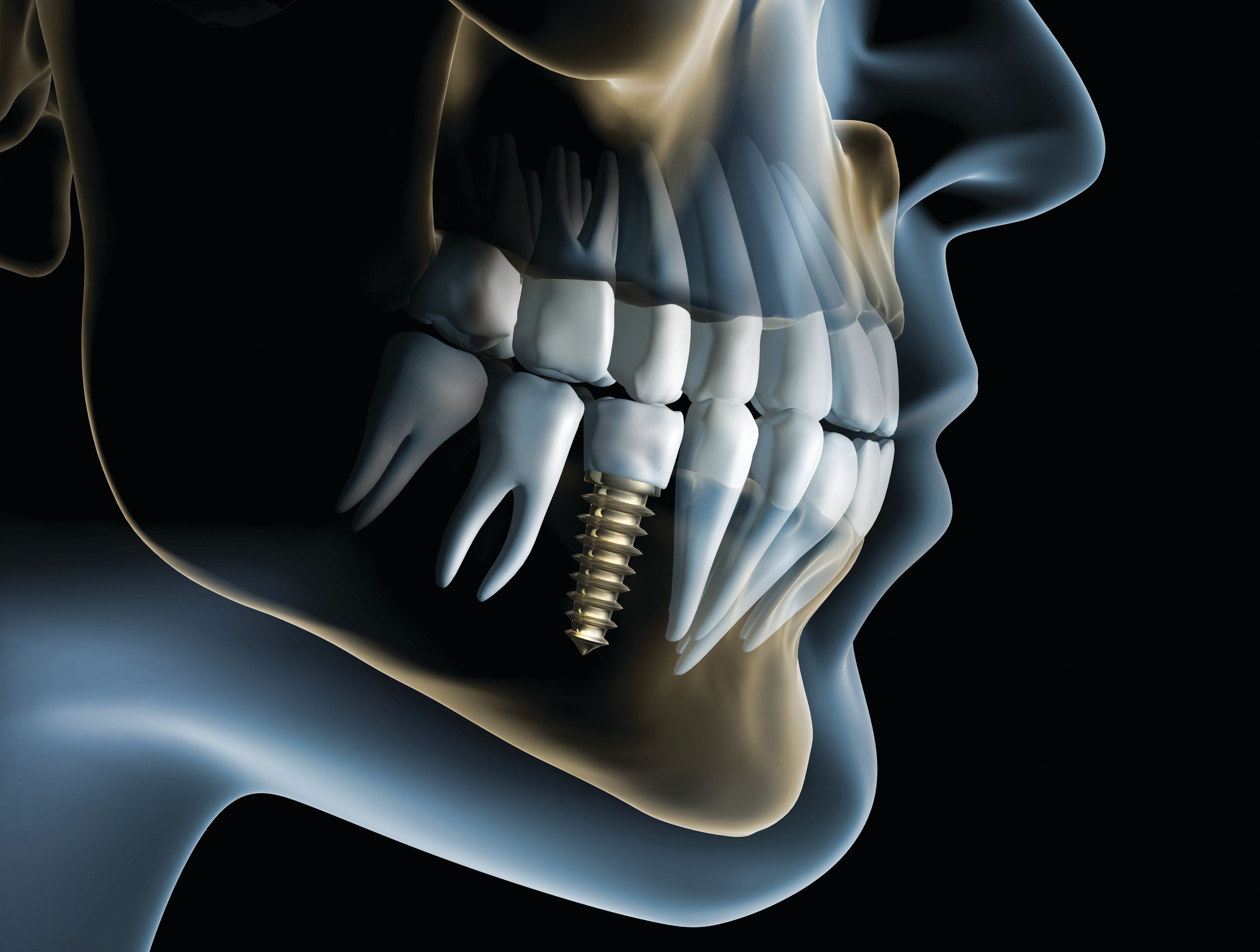
Osseointegration of anodized versus sandblasted implant surfaces in a guided bone regeneration acute dehiscence-type defect
This paper is an animal model study on minipigs, comparing the effect of crestal bone formation and osseointegration on different implant surfaces.
The authors noted some recent theories suggesting increased crestal bone loss around rough implant surfaces. The study was designed to assess crestal bone formation using three different implant surfaces.
The first was a novel gradient anodised (NGA) implant solution with a minimally rough surface at the collar (2mm), with a gradient increasing roughness beyond the neck of the implant. The NGA surface is hydrophilic and is commercially available.
The comparators were custom-cloned implants, copying the geometry of the NGA implant, with the first copy using a sandblasted and acid-etched hydrophilic surface. The second used a sandblasted and acid-etched hydrophobic surface. As such, both of these implants had a rough surface.
Histological analysis at two weeks showed no difference between the three implants in terms of crestal bone levels and new bone growth. However, at eight weeks, the first bone to implant contact and vertical bone creep were higher in the implants with a rough surface compared with the minimally rough surface.
The authors conclude that de novo crestal bone formation appears to be influenced by the surface roughness of the implant, with higher bone-to-implant contact and coronal bone growth on a rough surface within an animal model.
Shahdad S, Rawlinson S, Razaghi N, Patankar A, Patel M, Roccuzzo M, Gill T. Osseointegration of anodized versus sandblasted implant surfaces in a guided bone regeneration acute dehiscence-type defect: an in vivo experimental mandibular minipig model. Clin Oral Implants Res. 2025; 36(1): 127–141. https://doi.org/10.1111/clr.14369.
Read more