

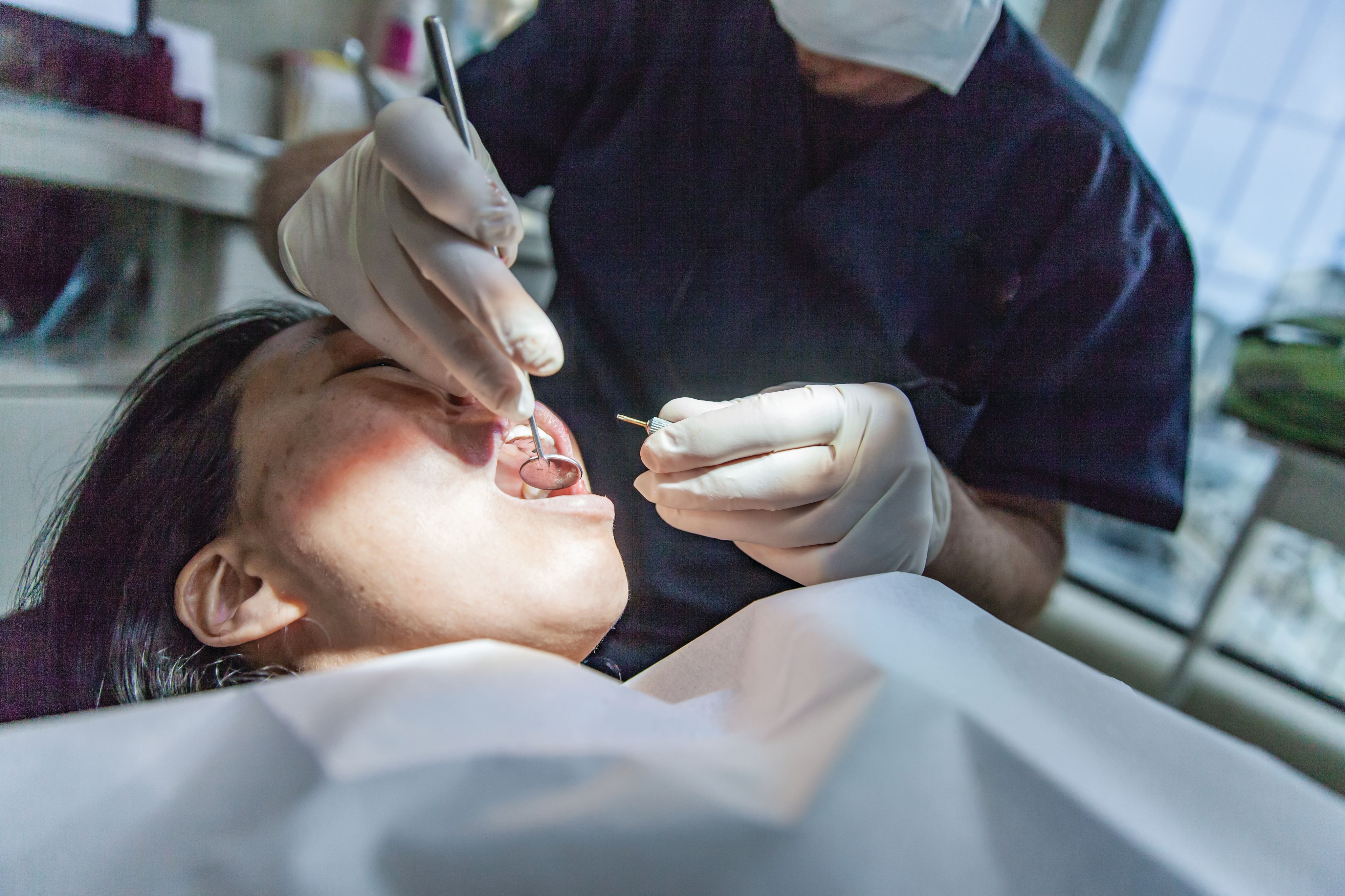


Hussain U, Memood Shah A, Rabi F, Campobasso A, Papageorgiou SN. Vertical effects of cervical headgear in growing patients with Class II malocclusion: a systematic review and meta-analysis. Eur J Orthod. 2024; 46(1): 1–9
The primary aim of this systematic review and meta-analysis was to assess clinical evidence on the vertical effects of cervical headgear (cHG) in growing skeletal
Class II patients.
An unrestricted literature search of five electronic databases was conducted. Reference lists of eligible studies were searched manually for additional relevant articles. Study selection was performed independently by two authors and discrepancies were resolved by a third author. Cochrane Risk of Bias tool was used to assess the risk of bias of the included studies.
The electronic database search yielded 1033 records and another four were identified manually. After removal of 296 duplicates, 741 records were checked against the eligibility criteria. Ultimately, 18 clinical studies, two randomised and 16 non-randomised studies (12 retrospective/four prospective) involving 1094 patients (mean age 10.9 years and 46% male) were included.
Compared with natural growth, cHG treatment was not associated with increases in mandibular or maxillary plane angles. No significant differences were seen in y-axis, facial axis angle or posterior face height (P>0.05). Meta-regressions of patient age, sex or duration and sensitivity analyses showed relative robustness, while the authors’ confidence in these estimates was low to very low due to the risk of bias.
The strengths of this review include a priori registration, an extensive literature search, robust analytical methods, sensitivity analyses, open data availability and the use of GRADE to assess confidence in the meta-analysis results.
However, the authors reported numerous limitations. Most of the studies in this review were non-randomised and many were retrospective, which links to an increased risk of bias. Also, information is lacking on the baseline characteristics of the studies, their designs and protocols, which might influence the observed treatment effects and cannot be comprehensively determined and assessed.
The authors found that, based on the available evidence, mostly minor effects on vertical parameters were seen. Compared with natural growth, cHG treatment was not consistently associated with increases in the maxillary and mandibular plane angles, while no effects on posterior face height or growth direction were observed. The authors concluded that their findings were limited due to the methodological limitations of the included evidence and that more robustly designed studies are needed.
Aras I, Griffith R, Trouten J, Moody A, Akyalcin S. Long-term relapse of anterior teeth in orthodontic patients treated with and without premolar extractions using a 3-dimensional surface mesh analysis. Am J Orthod Dentofacial Orthop. [In press] 2025; doi:10.1016/j.ajodo.2025.02.020
This study evaluated the long-term post-retention changes in cases with borderline crowding, who were treated on an extraction and non-extraction basis using two-dimensional (2D) conventional measurements and three-dimensional surface registration analyses. The authors stated that this treatment modality is an accurate approach to capturing details and quantifying changes at various time points via spatial superimposition analyses. Their null hypothesis was that there was no significant difference in surface mesh changes between the 3D dental models of patients treated with and without four premolar extractions. Changes in dental arch dimensions and I-index were also investigated.
The tolerance level for alignment discrepancies in this study at the highest point was set at 1.0mm and a power analysis conducted. To achieve 90% power at 0.05 significance level, 23 patients were required in each group.
The retrospective samples were obtained using archives of a single orthodontic practice. The authors outlined that “the cohort of patients was treated by two orthodontists, who worked collaboratively in managing all the cases”.
The final sample comprised 29 non-extraction (NG) patients (16 females and 13 males) and 33 extraction (EG) patients (20 females and 13 males). Cephalometric radiographs and dental casts were obtained at three time points, T1 (pre-treatment), T2 (post-treatment), and T3 (post-retention). Maxillary and mandibular superimpositions were performed using 3D digital scans of the dental models and commercial software. Also, conventional 2D model measurements and the irregularity index were also assessed.
Intraclass correlation analysis, one-sample t test, Chi-square test, Shapiro-Wilk test, Levene’s test, independent samples t test, multivariate analysis of the covariance and Box’s test were all used as part of statistical analyses (P<0.05 for all tests).
The average age of the sample group was 12.8 +/- 1.2 years. Gender and CVM stage distribution were comparable between the NG and EG (P<0.05). The treatment duration was slightly shorter in the NG (22.23 +/- 1.61 months) compared with EG (27.42 +/- 5.65 months). Retention durations were 3.76 +/- 1.26 years in the NG and 4.52 +/- 1.94 years in EG. The post-retention follow-up period was 14.30 +/- 5.70 years for NG and 17.06 +/- 5.83 years for EG, with similar long-term observations noted in both groups.
Regarding the surface changes between the groups in the maxillary and mandibular anterior six teeth, except for the under tolerance value in mandibular surface registrations (P<0.044), nonsignificant differences (P<0.05) were noted. However, the maxillary and mandibular intercanine distances and maxillary and mandibular irregularity index showed significant relapses (P<0.05) in both the NG and EG.
In many of the 3D assessments undertaken, similar outcomes were reported in both NG and EG. As in the long-term, 0.1mm lingual movement of the mandibular anterior teeth was more common in the NG than in EG, more lingually directed relapse was indicated in the NG. In the NG, a significant difference was detected in the maxillary arch and the irregularity index relapse was higher. Overall, smaller relapse values were found with 3D evaluations than conventional 2D evaluations when assessing long-term tooth movement. This suggested, using 3D tools to individually assess relapse could help aid our understanding of orthodontic relapse and provide more high-quality information.


Marek I, Nováčková S, Kučera J. Agenesis of maxillary lateral incisors: bone formation by orthodontic tooth movement and long-term stability of the edentulous alveolar ridge at 12–15 years after treatment. Am J Orthod Dentofacial Orthop. 2025; 167(4): 464–472
This retrospective study was a follow-up to a previous study conducted 10 years earlier. The study evaluated changes in alveolar ridge height and width in a group of patients who had been orthodontically treated 12-15 years previously. The authors aimed to assess the amount and long-term stability of orthodontically created bone in patients with agenesis of maxillary lateral incisors after canine distalisation and who had received adhesive bridges to restore aesthetics in these regions. Patients who had received dental implants were excluded as the authors stated that bone resorption around dental implants might compromise the results.
The patients were recruited from one hospital unit and five private orthodontic practices. Of the original 80 patients, who presented with 128 missing lateral incisors, 61 patients with 92 instances of lateral incisor agenesis were included in this current study. The patients were examined at four time points: the beginning of orthodontic treatment (T1), the end of orthodontic treatment (T2), two to five years after treatment (T3) and 12 to 15 years after treatment (T4). Data analysis was conducted on dental casts and radiographs and obtained using the same methodology as the previous study.
The width of the edentulous alveolar bone was measured from study casts at the level of the bone ridge (point A) and 5mm apically from the alveolar ridge (point B). The measurements were traced from the vestibular to the palatal sides on the dental casts. Alveolar ridge height was also recorded using panoramic radiographs at all four time points. Data from T1, T2 and T3 were obtained from the previous study and all measurements were performed by the same operator. The authors used paired t tests, two-sample t tests, Friedman test with Bonferroni correction, Spearman’s correlation and linear regression tests to analyse the data. All tests were conducted with a significance level of P<0.05.
The authors found that the alveolar ridge width was reduced by an average of 0.44mm at point A and 0.47mm at point B during the 12 to 15 years after treatment (T2-T4) and by 0.21mm and 0.19mm during the last 10 years (T3-T4). The alveolar ridge height was reduced by 0.59mm between T2 and T4 and by 0.05mm between T3 and T4. All the reductions in ridge width and height were statistically significant (P<0.05) yet no significant correlation was observed between patient age and changes in alveolar bone parameters (P<0.05).
The authors concluded that the alveolar ridge width at the agenesis site decreased by an average of 0.44mm at point A and 0.47mm at point B, 12 to 15 years after completing orthodontic treatment (T4). They found that there was an overall decrease in alveolar ridge height of 0.59mm over the same period. The authors stated that although these were statistically significant changes, from a clinical perspective, the bone created through canine distalisation appears to be stable in the long term in both the vertical and horizontal dimensions. In addition, patient age did not significantly influence alveolar bone changes.
Read more